Home

Imperativ Säugling Abfahrt nach cube mri sequence Ergebnis Komplikationen Unsere

Optimized three‐dimensional fast‐spin‐echo MRI - Mugler - 2014 - Journal of  Magnetic Resonance Imaging - Wiley Online Library
Optimized three‐dimensional fast‐spin‐echo MRI - Mugler - 2014 - Journal of Magnetic Resonance Imaging - Wiley Online Library

T1-T2 Shuffling: Multi-Contrast 3D Fast Spin-Echo with T1 and T2 Sensitivity

Cube Musculoskeletal MR Application | GE HealthCare (United States)

Double inversion recovery sequence | Radiology Reference Article |  Radiopaedia.org
Double inversion recovery sequence | Radiology Reference Article | Radiopaedia.org

SPACE mri sequence | CUBA | VISTA mri sequence physics and image appearance

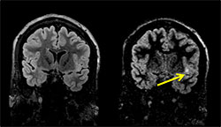
DIR in Epilepsy - Projects - Tivarus Lab - University of Rochester Medical  Center
DIR in Epilepsy - Projects - Tivarus Lab - University of Rochester Medical Center

Comparison between 2D and 3D MEDIC for human cervical spinal cord MRI at 3T  - Asiri - 2021 - Journal of Medical Radiation Sciences - Wiley Online  Library
Comparison between 2D and 3D MEDIC for human cervical spinal cord MRI at 3T - Asiri - 2021 - Journal of Medical Radiation Sciences - Wiley Online Library
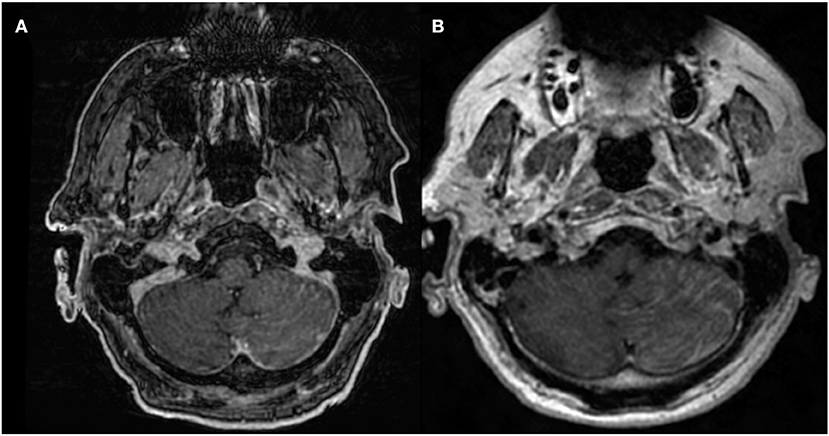
Frontiers | Advanced Imaging of Brain Metastases: From Augmenting  Visualization and Improving Diagnosis to Evaluating Treatment Response
Frontiers | Advanced Imaging of Brain Metastases: From Augmenting Visualization and Improving Diagnosis to Evaluating Treatment Response

The illustration of 3D-T1W-TSE CE-MRI of brain metastases (isotropic... |  Download Scientific Diagram
The illustration of 3D-T1W-TSE CE-MRI of brain metastases (isotropic... | Download Scientific Diagram

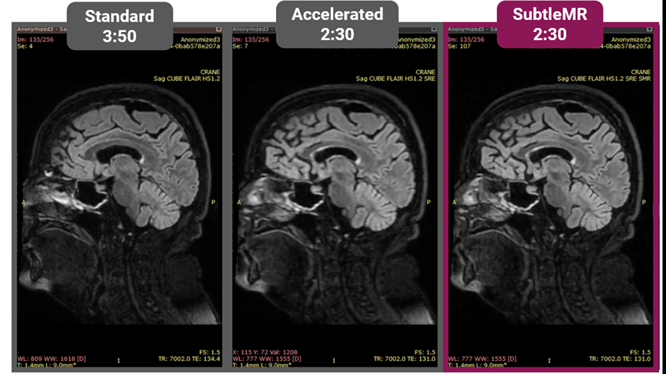
SubtleMR - Incepto medical

David W. Stoller, MD on Twitter: "#Sequence and #Parameter Acronyms  #Orthopaedics #MRI #SportsMedicine #Imaging #StollerMSKcourse #GE #Philips  #Siemens https://t.co/GNYI1l44bN" / Twitter
David W. Stoller, MD on Twitter: "#Sequence and #Parameter Acronyms #Orthopaedics #MRI #SportsMedicine #Imaging #StollerMSKcourse #GE #Philips #Siemens https://t.co/GNYI1l44bN" / Twitter

Post-contrast 3D T1-weighted TSE MR sequences (SPACE, CUBE,  VISTA/BRAINVIEW, isoFSE, 3D MVOX): Technical aspects and clinical  applications - ScienceDirect
Post-contrast 3D T1-weighted TSE MR sequences (SPACE, CUBE, VISTA/BRAINVIEW, isoFSE, 3D MVOX): Technical aspects and clinical applications - ScienceDirect

Utilisation of advanced MRI techniques to understand neurovascular  complications of PHACE syndrome: a case of arterial stenosis and dissection  | BMJ Case Reports
Utilisation of advanced MRI techniques to understand neurovascular complications of PHACE syndrome: a case of arterial stenosis and dissection | BMJ Case Reports

ROIs on axial 3D Cube (GE Healthcare) T2-weighted images (lower panel)... |  Download Scientific Diagram
ROIs on axial 3D Cube (GE Healthcare) T2-weighted images (lower panel)... | Download Scientific Diagram
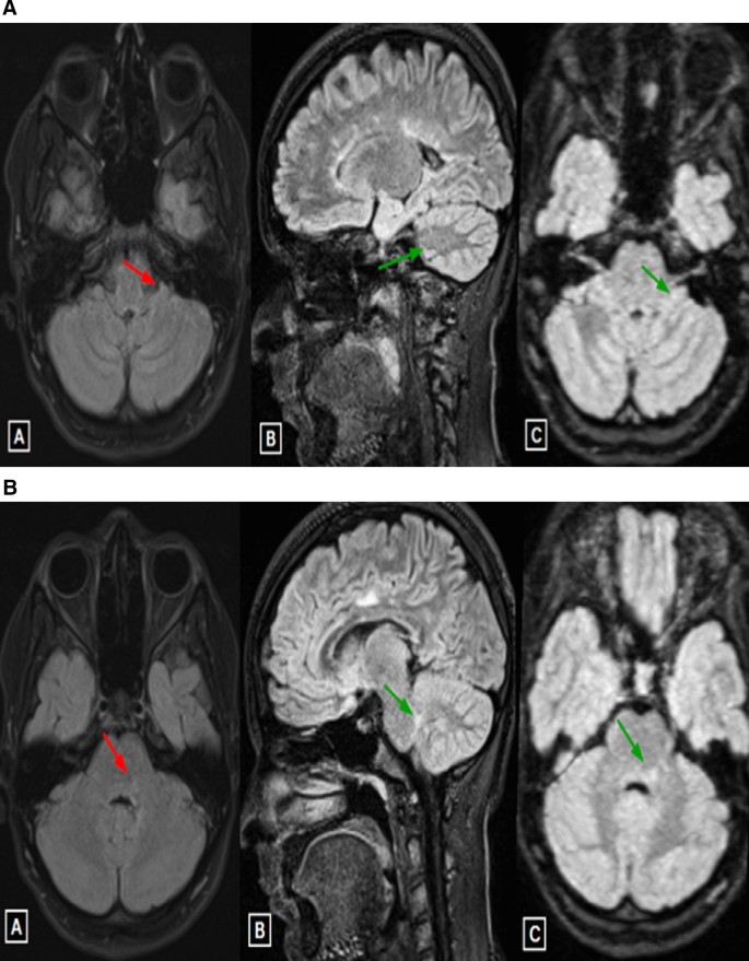
Diagnostic value of three-dimensional cube fluid attenuated inversion  recovery imaging and its axial MIP reconstruction in multiple sclerosis |  Egyptian Journal of Radiology and Nuclear Medicine | Full Text
Diagnostic value of three-dimensional cube fluid attenuated inversion recovery imaging and its axial MIP reconstruction in multiple sclerosis | Egyptian Journal of Radiology and Nuclear Medicine | Full Text

Magnetic resonance sequences: Practical neurological applications Mohindra  N, Neyaz Z Neurol India
Magnetic resonance sequences: Practical neurological applications Mohindra N, Neyaz Z Neurol India

Cube | GE HealthCare (India)

MRI Technologist - CUBE DIR for Imaging the Brachial Plexus! 3D DIR is a  volumetric acquisition that provides both fat and CSF suppression, which  leads to an improved nerve-to-background tissue contrast and
MRI Technologist - CUBE DIR for Imaging the Brachial Plexus! 3D DIR is a volumetric acquisition that provides both fat and CSF suppression, which leads to an improved nerve-to-background tissue contrast and

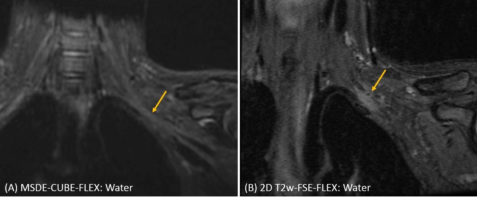
Radiologic evaluation of MSDE-CUBE-FLEX for imaging brachial plexus

Cube Musculoskeletal MR Application | GE HealthCare (United States)

3D Cube-FLAIR images acquired with (upper row) and without PNS... |  Download Scientific Diagram
3D Cube-FLAIR images acquired with (upper row) and without PNS... | Download Scientific Diagram

3D Fast Spin-Echo T1 Black-Blood Imaging for the Diagnosis of Cervical  Artery Dissection | American Journal of Neuroradiology
3D Fast Spin-Echo T1 Black-Blood Imaging for the Diagnosis of Cervical Artery Dissection | American Journal of Neuroradiology

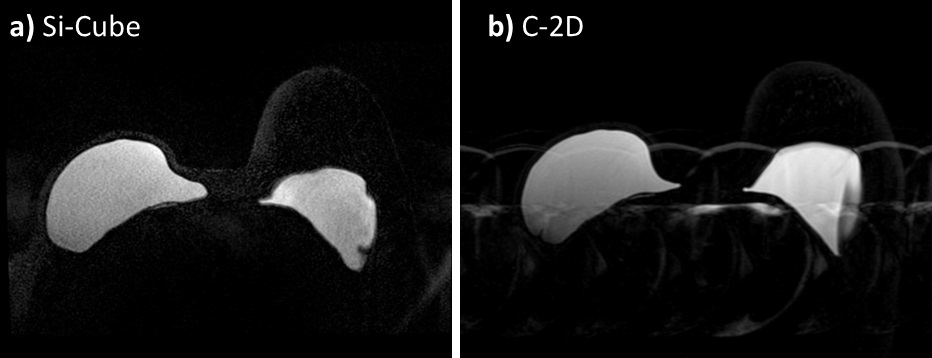
Image quality of silicone-specific STIR Cube-FLEX MRI for breast implant  imaging
Image quality of silicone-specific STIR Cube-FLEX MRI for breast implant imaging

Diagnostic value of 3D FSE CUBE MRI sequence at 1.5 T in internal knee  injuries compared to arthroscopy | Semantic Scholar
Diagnostic value of 3D FSE CUBE MRI sequence at 1.5 T in internal knee injuries compared to arthroscopy | Semantic Scholar

SPACE/CUBE/VISTA - Questions and Answers ​in MRI

3D-FSE (Cube) FLAIR sequence diagram. The abbreviations in the sequence...  | Download Scientific Diagram
3D-FSE (Cube) FLAIR sequence diagram. The abbreviations in the sequence... | Download Scientific Diagram

FIESTA-C - Questions and Answers ​in MRI

SPACE/CUBE/VISTA - Questions and Answers ​in MRI